Hypopigmented mycosis fungoides
Clinical features
•Associated with a good prognosis
•More commonly seen in children & adolescents although any age can be affected
•M=F
•Predilection for African & Asian descent; much less often in Caucasians
•Generally asymptomatic, scaly, hypopigmented patches on trunk, buttocks & extremities
•Clinically mistaken for vitiligo, post inflammatory hypopigmentation, tinea versicolor & pityriasis alba
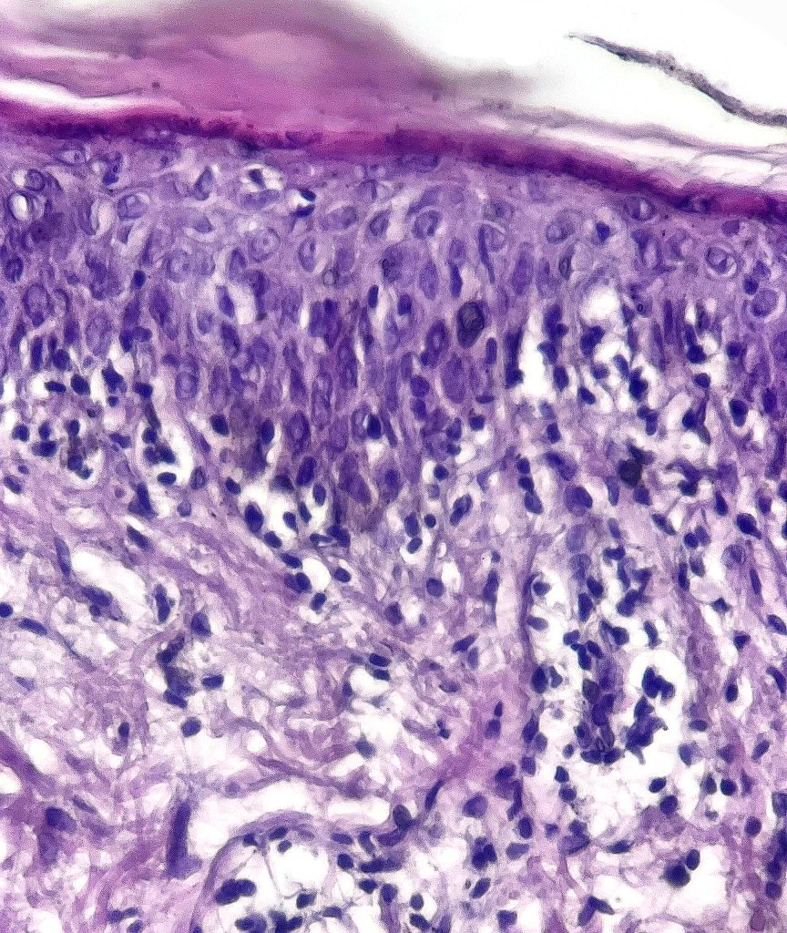
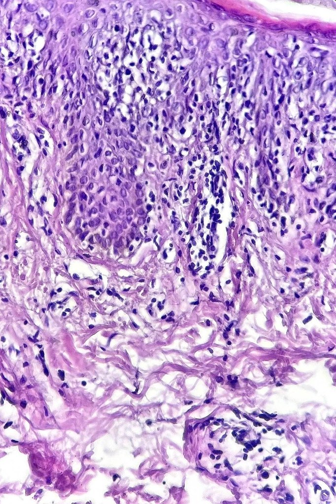
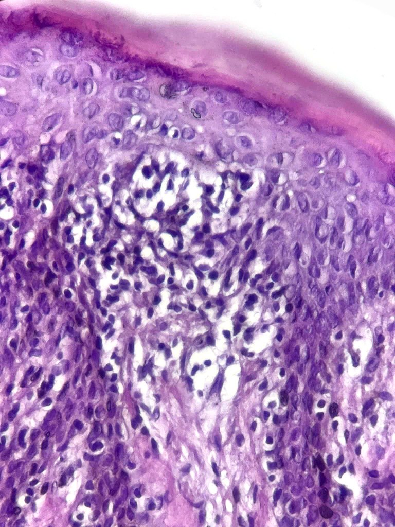
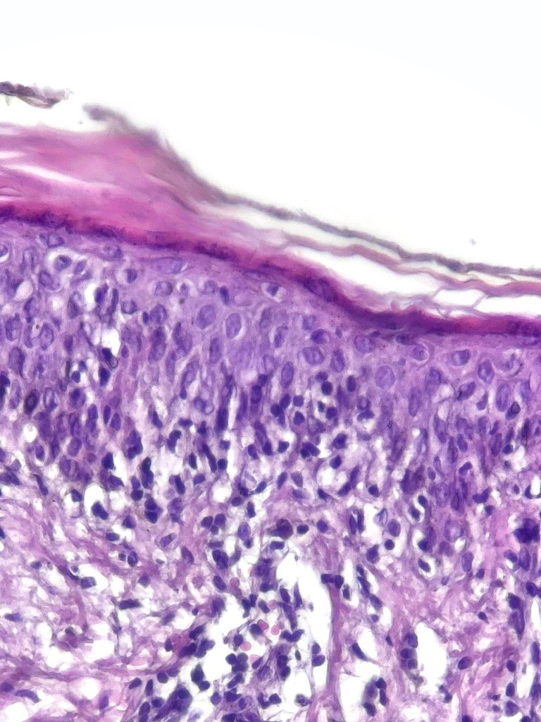
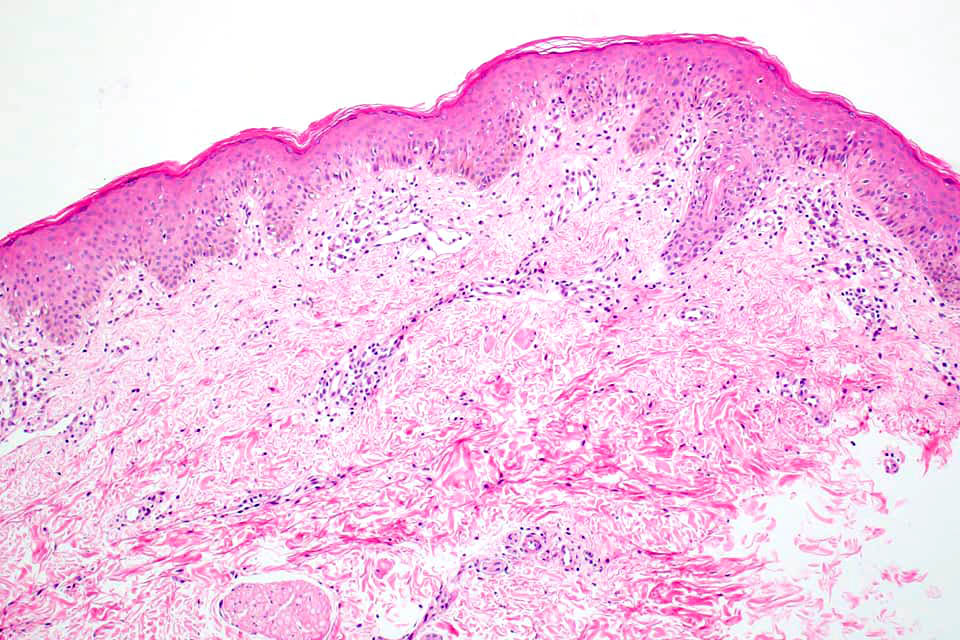
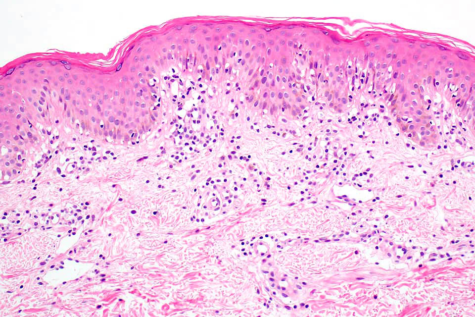
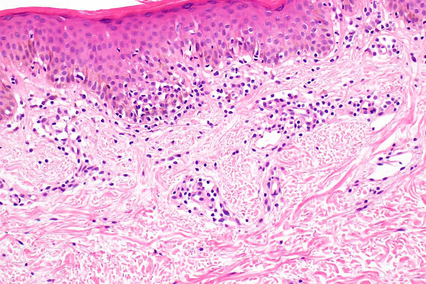
Histological features
•Indistinguishable from classical mycosis fungoides with the additional feature of marked pigmentary incontinence
•CD8+ve cells may predominate
•Hypopigmentation likely associated with melanocytic toxicity & reduced melanocyte population although this isn’t always a feature
•Masson-Fontana may show reduced melanin with SOX10 & with Melan-A showing reduced numbers of melanocytes
Childhood and adolescent mycosis fungoides
•Exceedingly rare
•Lesions are frequently hypopigmented
•Presents with patch & plaque stage disease
•Prognosis is generally good
•Histology is identical to classical mycosis fungoides
•A CD8 immunophenotype is common particularly in hypopigmented variants


Leave a comment