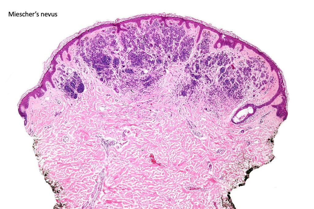
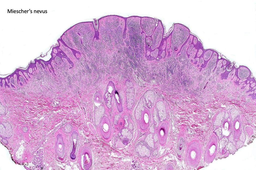
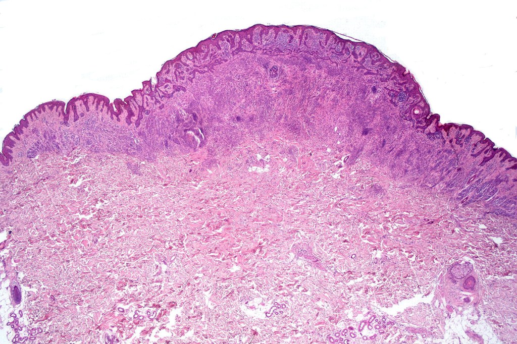
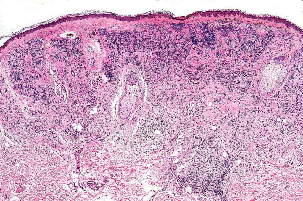
•The late Bernie Ackerman classified acquired melanocytic nevi into Miescher nevus which most commonly occurs on the face and Unna nevus which generally presents on or below the neck
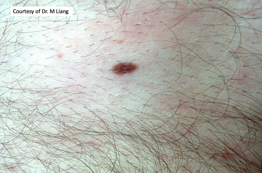
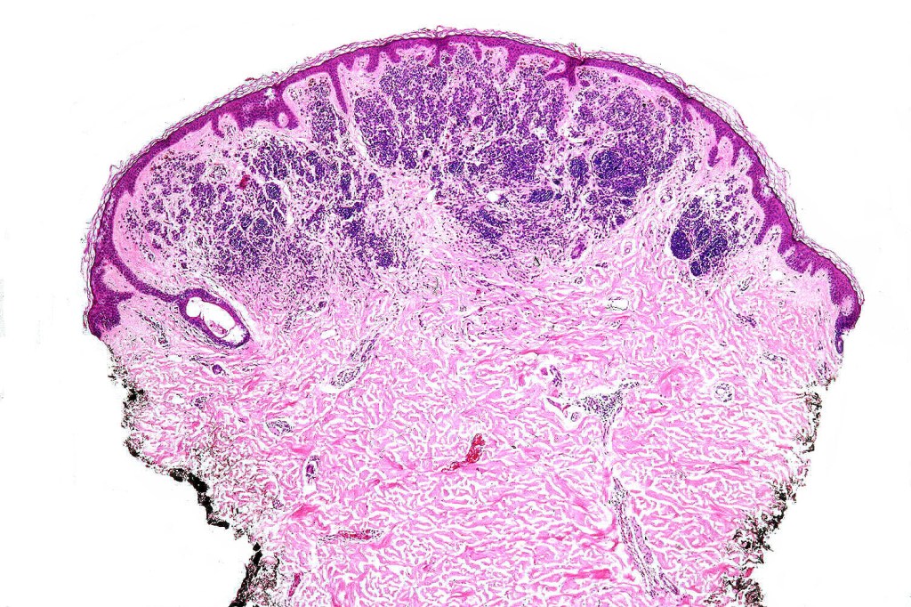
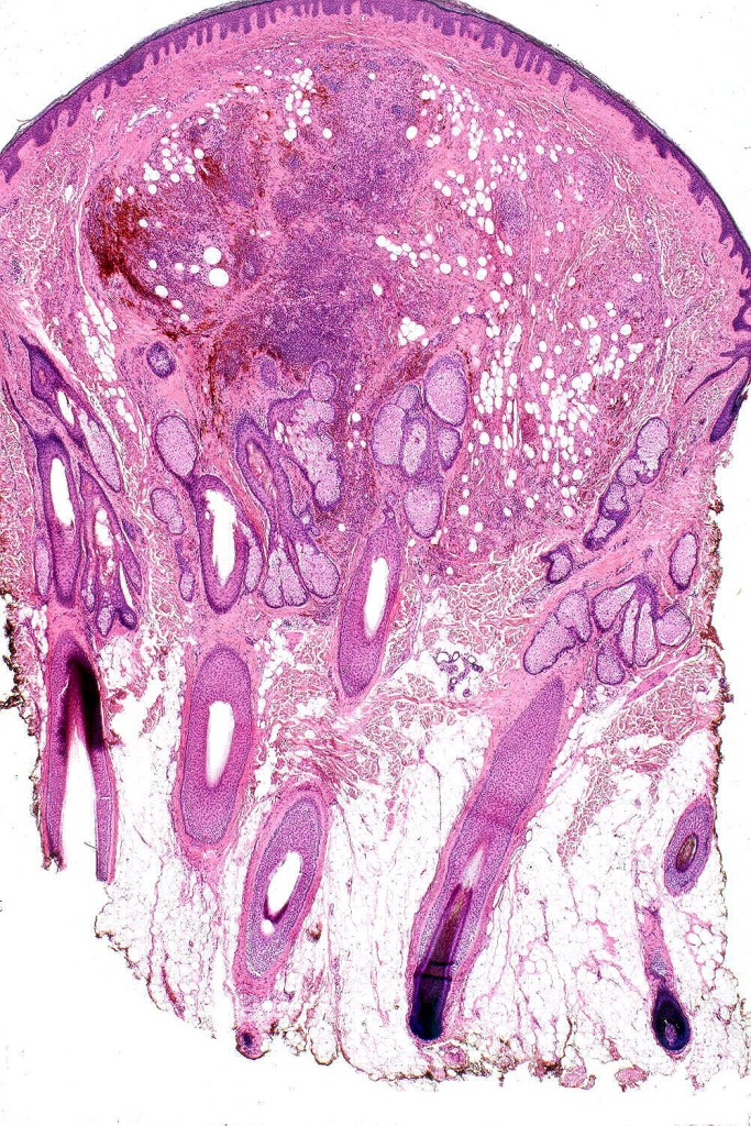
•Miescher nevus is typically dome-shaped
•In Miescher nevus, the nevus is generally dermal, wedge shaped & extends into the reticular dermis
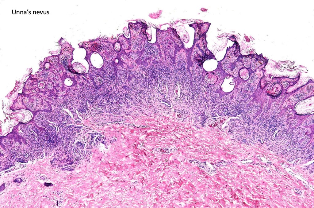
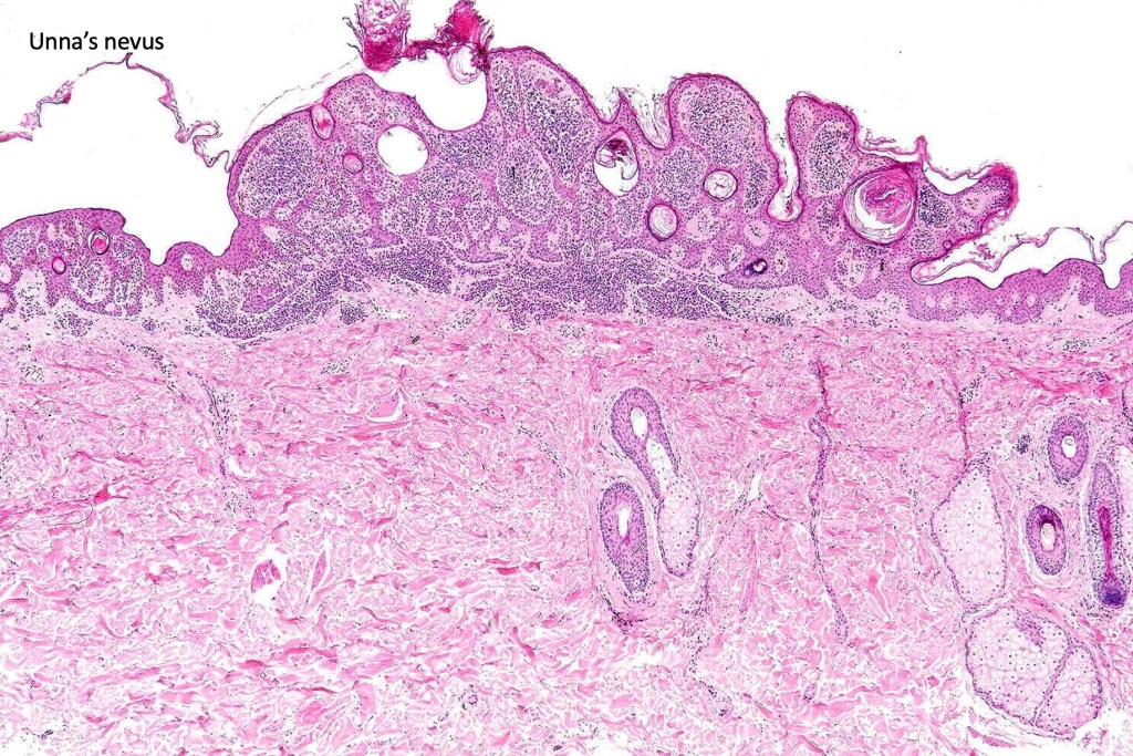
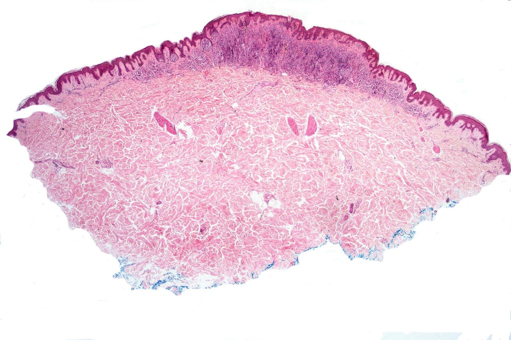
•Unna nevus presents as an exophytic, light to dark brown, polypoid, verrucous or sometimes sessile lesion with a predilection for the neck, trunk & arms
•In Unna nevus, the nevus is predominantly adventitial & therefore is restricted to the papillary dermis & and often includes a perifollicular adventitial element
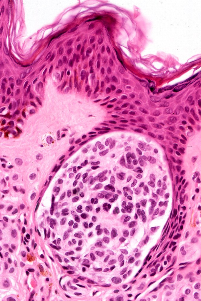
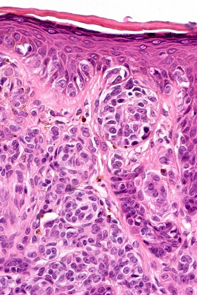
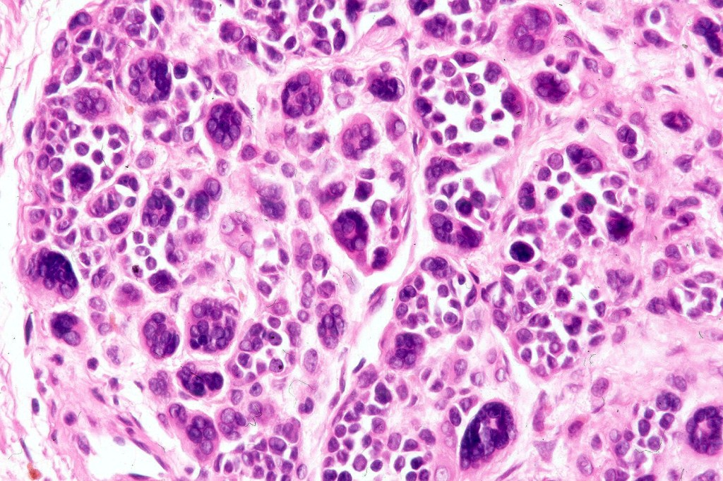
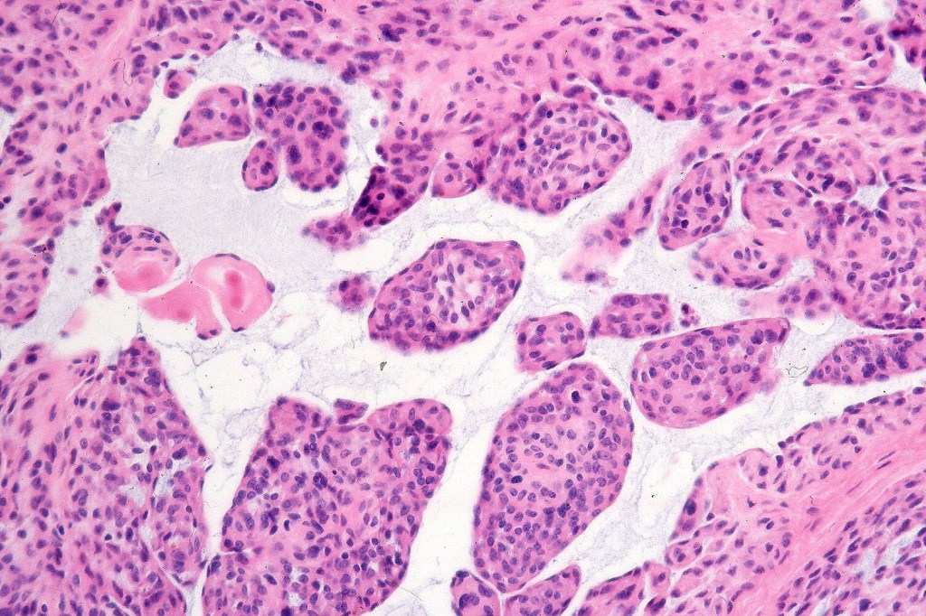
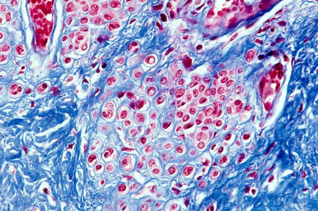
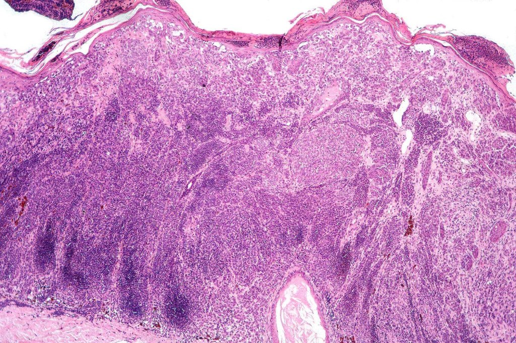
Type-A cells
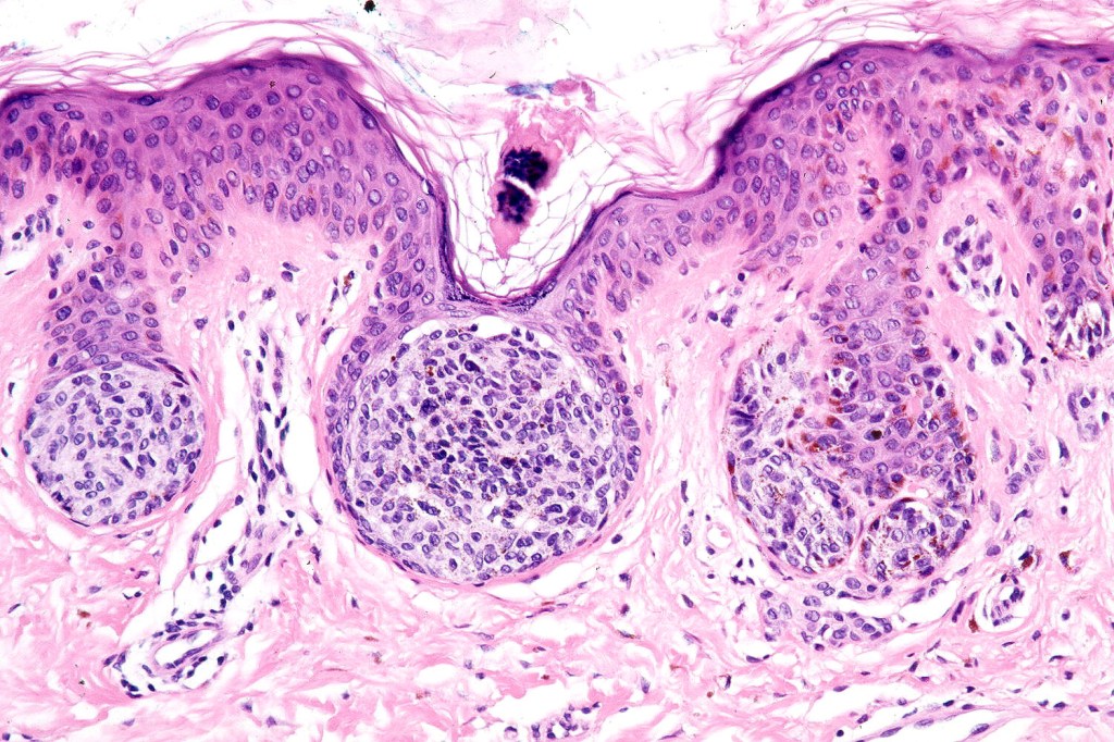
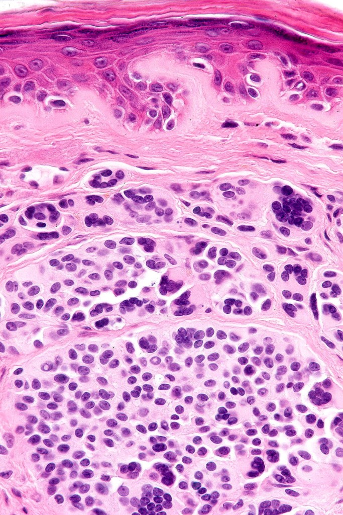
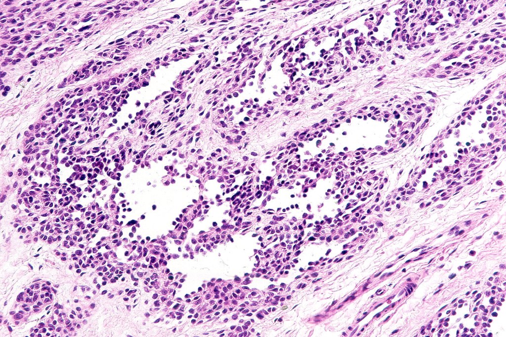
Junctional nevus

.Nests are prefernatially located at the tips of the rete ridges (compare with dysplastic where nests are often present on the sides of the rete and overling the dermal papilla)
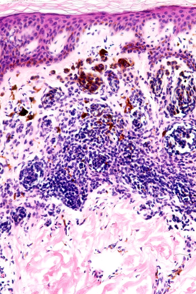
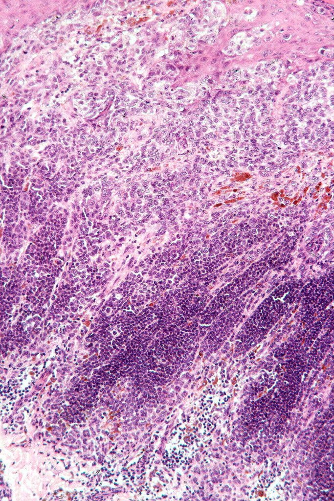
.Composed of type-A cells- abundant eosinophilic or finely granular, pigmented cytoplasm
.Vesicular nuclei with small nucleoli
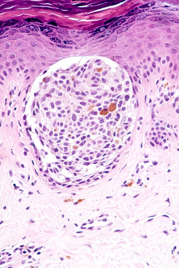
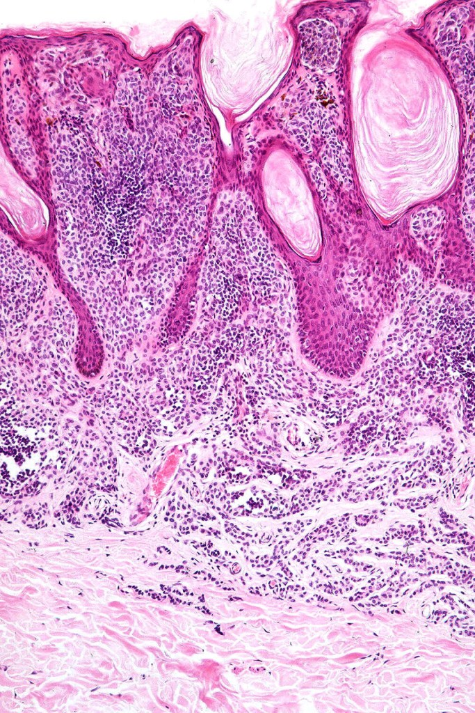
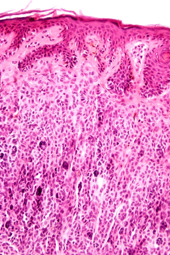
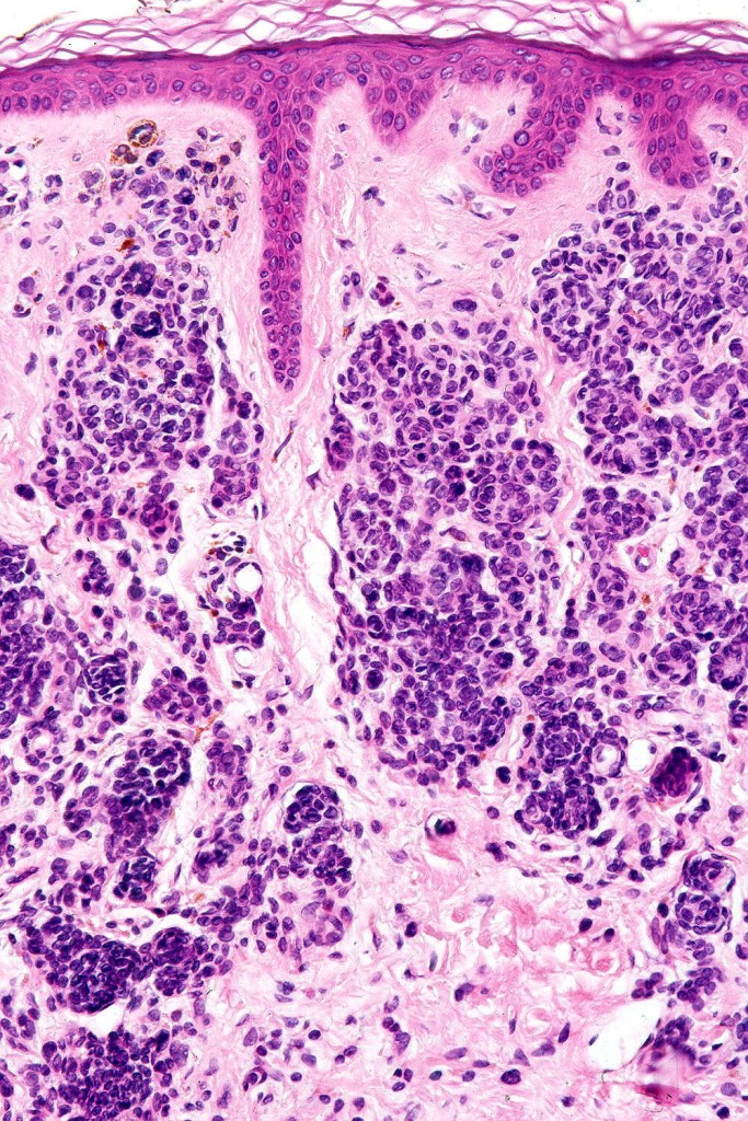
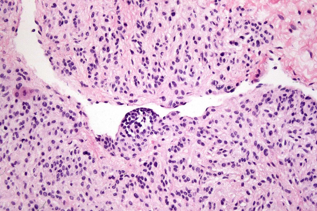
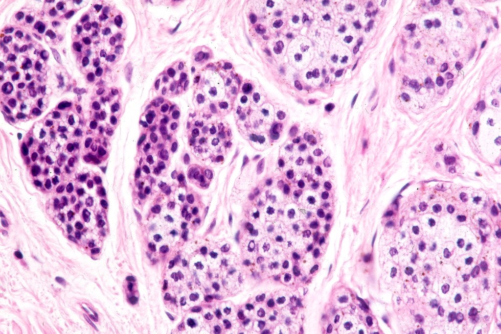
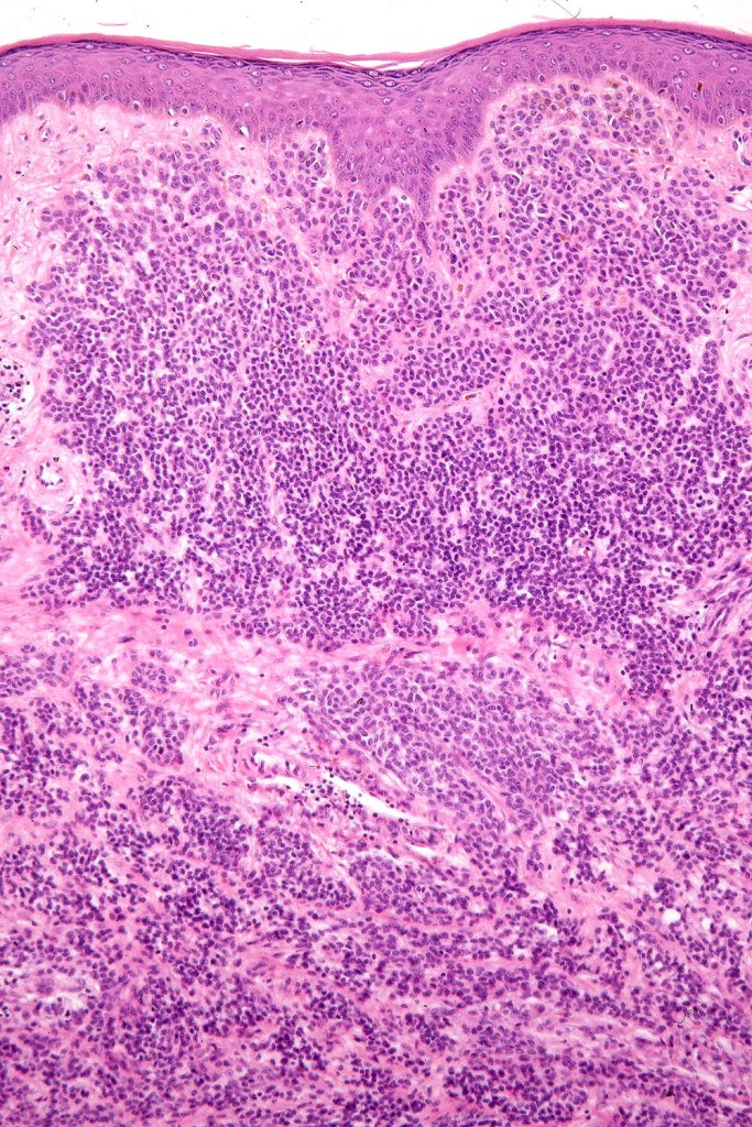
Compound nevus
The nevus includes junctional and dermal components (the latter often comprises type-A cells superficially merging wiht type-B nevus cells in the deeper reaches)
.Type-B nevus cells have scant cytoplasm and dense, basophilic cyoplasm
.The nevus shows maturation with depth- nest and cell size get smaller with increasing depth
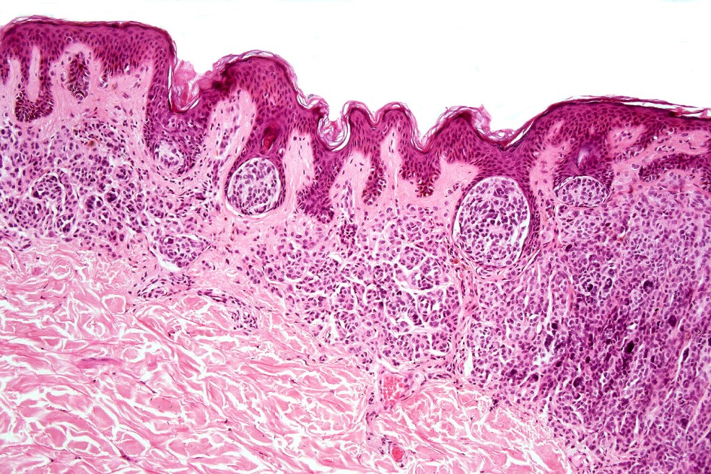
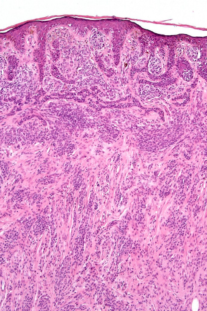
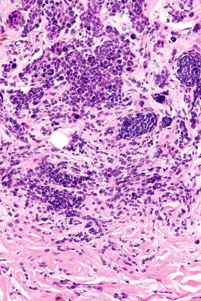
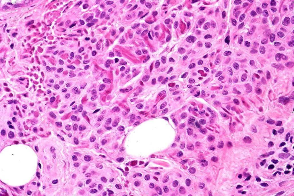
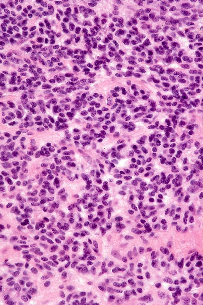
Dermal nevus
.The nevus is confined to the papillary dermis with or without involvement of the reticular dermis
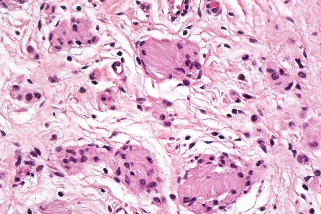
.Superficially may be composed of type-A nevus cells or just type-B nevus cells. In the deeper reaches, there are type-C spindled cells sometimes showing neurotization and forming Meissner corpuscle-like bodies
. Multinucleate giant cells are sometimes present
.Adipocytes can be present
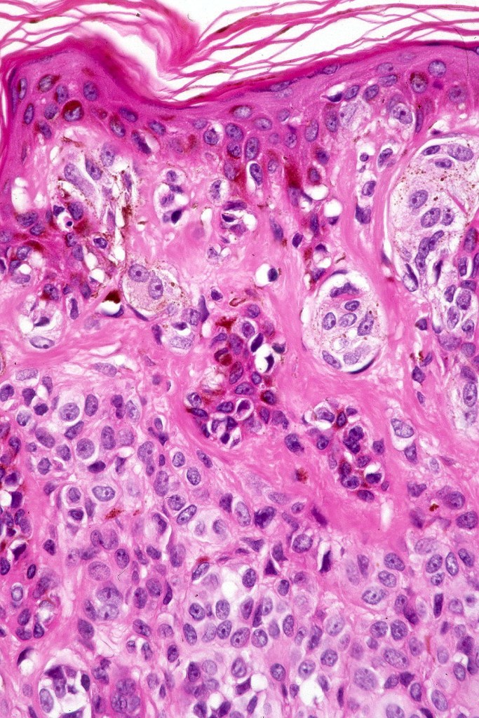
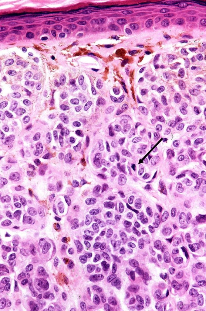
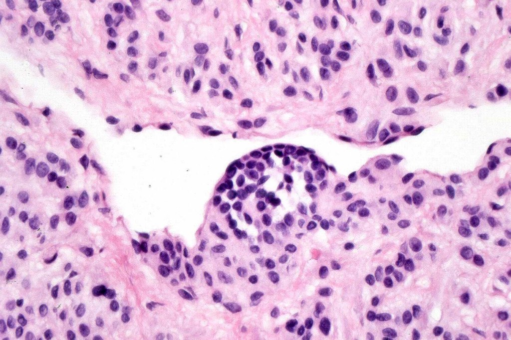
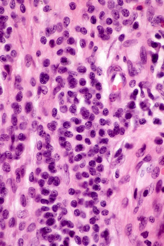
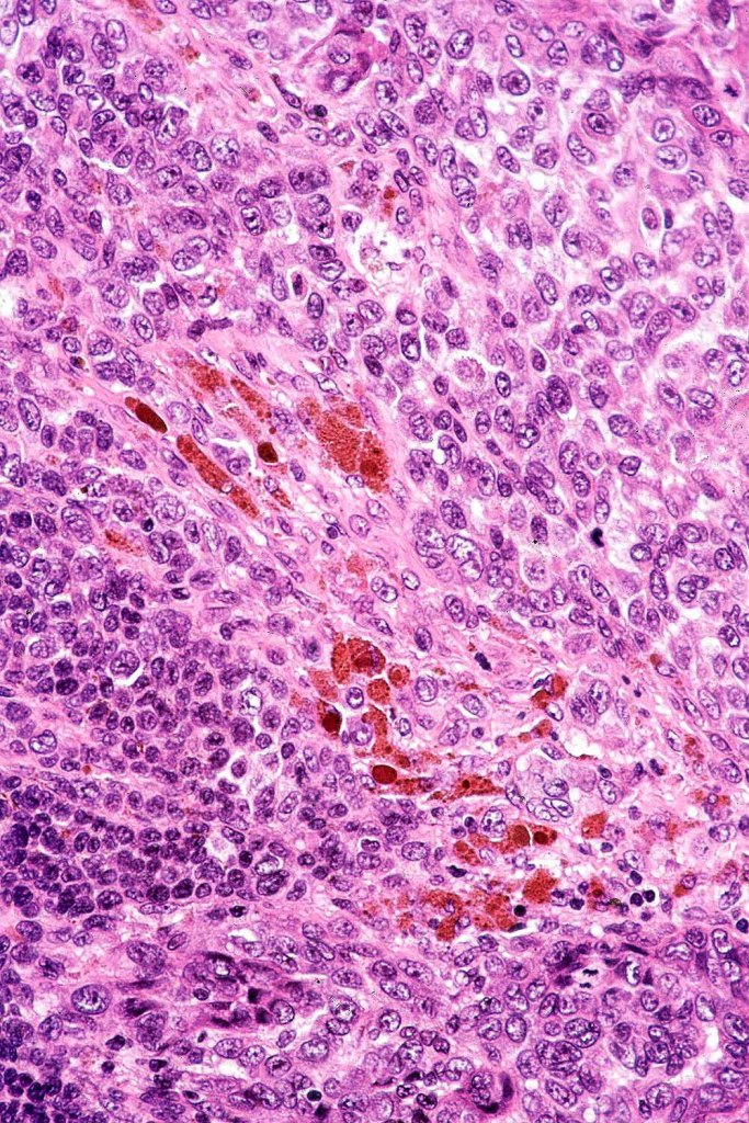
Type-A nevus cells with mitosis

Leave a comment