Clinical features
•Rare & may present in the dermis, subcutaneous fat or soft tissue
•Cutaneous lesions present mostly in adolescents & young adults with a predilection for males
•Firm to hard nodule 0.5-2.5 cm
•Age range newborn-93 years
•Soft tissue tumors are often much larger & present most often limbs & limb girdles with an equal sex incidence
•Malignant myoepithelioma in the soft tissues is exceedingly rare and has a 40-50% risk of local recurrence, metastasis rate of 30-50% & high mortality
Histological features
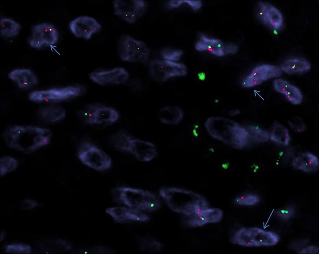
•Rearrangement of EWSR1 in syncytial & soft tissue tumors
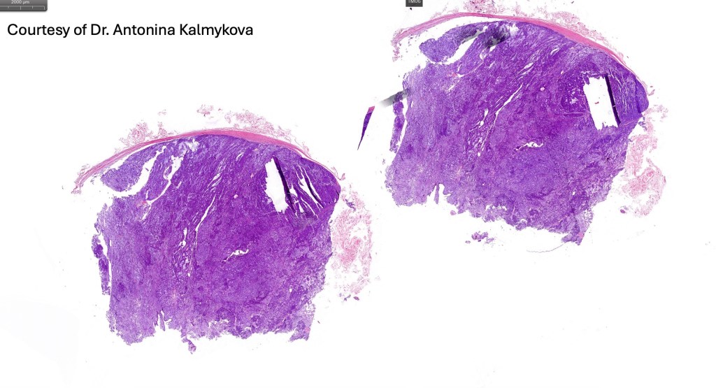
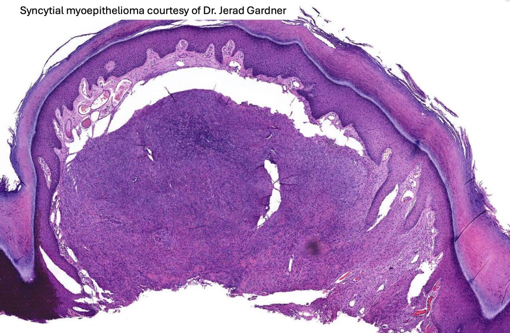
•The epidermis may show a collarette surrounding an often dome-shaped nodule
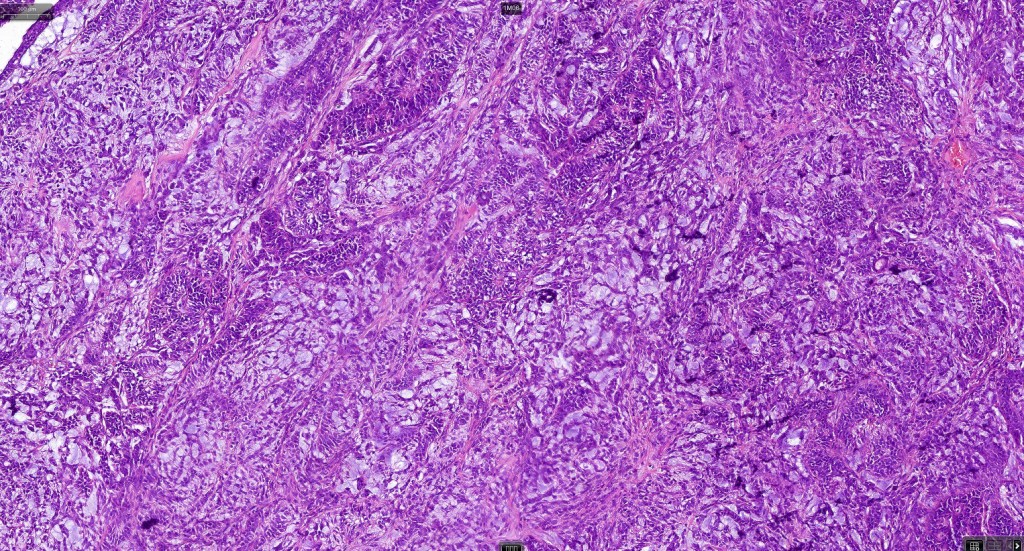
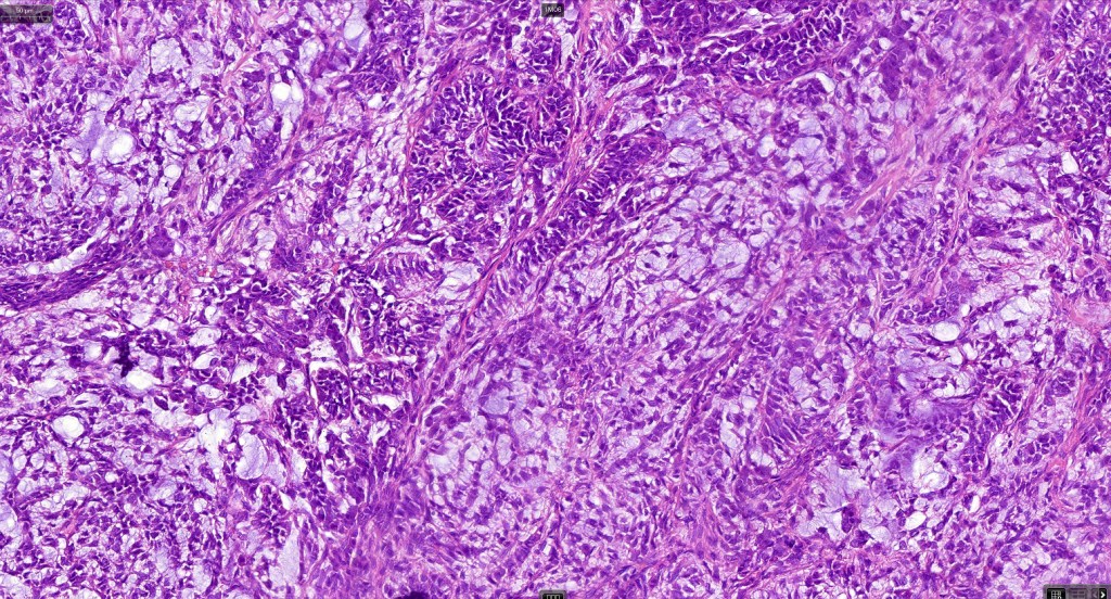
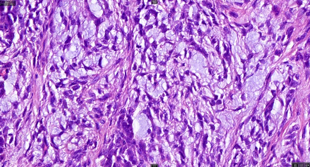
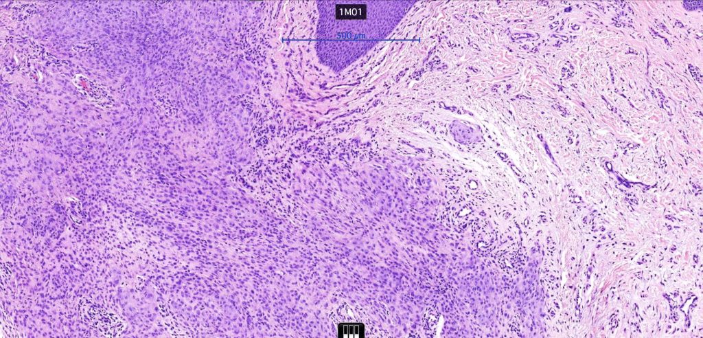
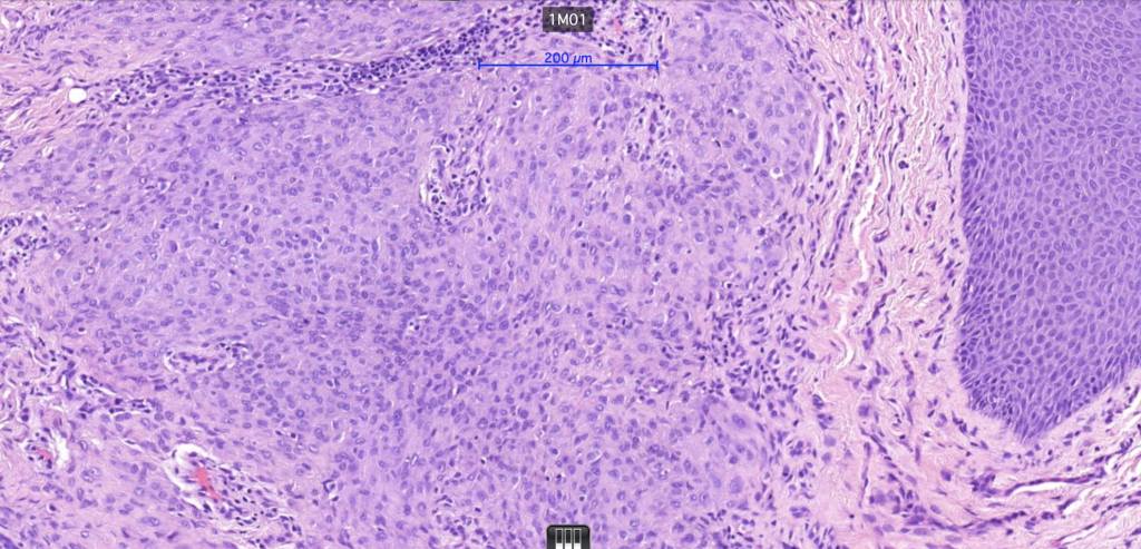
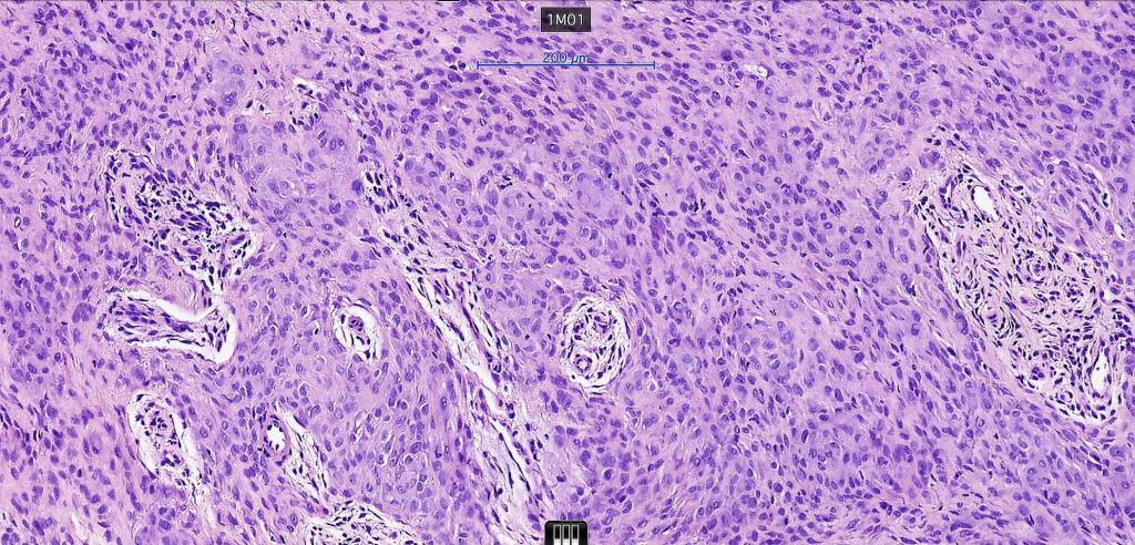
•Pure population of myoepithelial cells dispersed in sheet-like, reticular, whorled or fascicular patterns in a myxoid or hyaline stroma
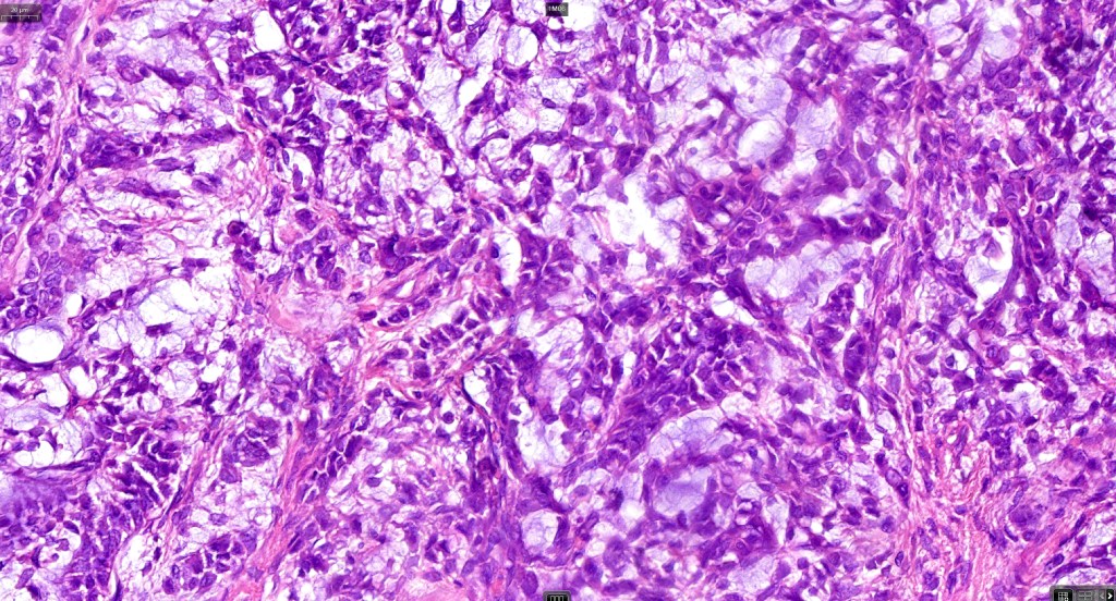
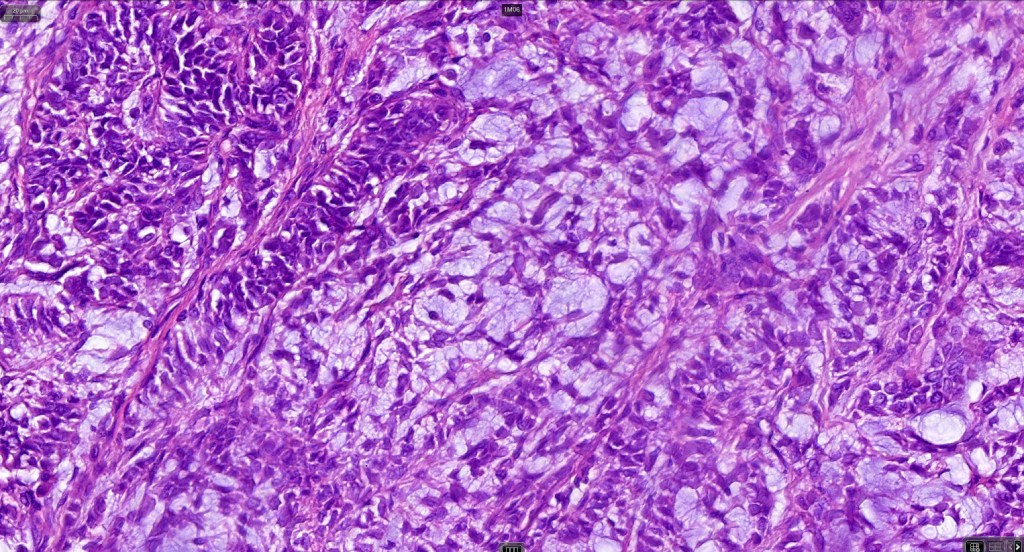
•Cell types include epithelioid, spindled, histiocytoid & plasmacytoid
•Oncocytic & clear cell sometimes present
•No glandular or ductal differentiation
•No pleomorphism and absent or scanty mitoses
•Exceptionally, chondroid, osteoid or adipocytes
•Collagenous crystalloid inclusions sometimes evident
•Pigmented variant
•Mucinous variant
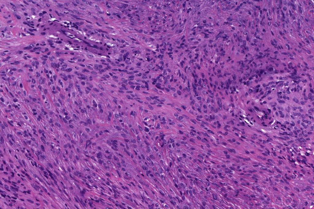
•Syncytial myoepithelioma characterized by sheet-like growth of ovoid to spindle cells with pale cytoplasm, syncytial borders & vesicular nuclei
•In malignant myoepithelioma there is an infiltrating border, marked pleomorphism, high mitotic rate & necrosis. Perineural infiltration & lymphovascular invasion may be seen
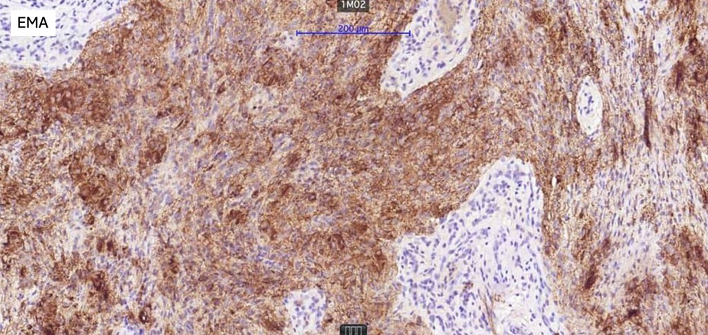
•AE1/AE3, GFAP, S100, SOX10, EMA,
•Variable p63, SMA & calponin +ve
.Syncytial myoepithelioma EMA & S100 +ve


Leave a comment